Stephen Ball Malvern Instruments Nanoparticle and Molecular Characterization Product Marketing Manager
Lead
After ten years of investment, nanotechnology has matured. Nanomedical materials are now emerging in clinical and medical practice. From a business perspective, by the end of 2015, this carefully cultivated research is expected to make the value of the biomedical nanotechnology market exceed 70 billion US dollars. In practical terms, this means that the methods of disease targeting and treatment may change.
Nanoscale colloidal gold has great potential in multiple therapeutic and biotechnology applications. Taking drug delivery as an example, by controlling the unique chemical, physical, and electronic properties of colloidal gold, researchers can develop drug-nanoparticle complexes that target drug delivery to enhance specific biological targets such as diseased tissues and cancer cells. Biodistribution and pharmacokinetics. Therefore, gold nanoparticles have become an important platform for innovating intracellular delivery media and controlling the particle size of nanoparticles in the formulation process, and the definition of this function is crucial.
This article describes the importance of detecting particle size in biomedical nanotechnology and demonstrates how to measure nanoscale and sub-nanoscale colloidal gold using advanced dynamic light scattering (DLS) techniques.
Flashing Things: A Brief History of Gold Therapy
Since ancient times, people have thought that gold has therapeutic properties. However, until the 18th century, the antibacterial properties of cyanide-containing gold salts were discovered. Time flies, and today, more than a hundred years later, gold salt has been used for routine treatment and control of rheumatoid arthritis. But beyond these traditional treatments, modern people have ignited new interest in the colloidal form of gold.
Many of the properties of colloidal gold make it ideal for clinical applications in a variety of nanomaterial matrices. Its chemical and physical inertness ensures that it is safe and non-toxic in vivo, while the fine particle size allows it to pass through the cell membrane without damage to the cells. In addition, gold nanoparticles suspended in an aqueous medium form negatively charged ions, and have strong affinity for biological macromolecules such as proteins and antibodies, so that they form bioligands around the ions. Currently, various biomedical applications such as imaging probes, diagnostic agents, and advanced drug delivery are developing these unique physical properties. In the field of advanced drug delivery, research and development activities using gold nanoparticles to provide cutting-edge delivery mechanisms for a range of traditional and innovative therapies for oral insulin, advanced anti-cancer drugs and DNA complexes required for advanced gene therapy are also underway. .
As with all microparticle therapies, pharmacokinetic properties such as bioavailability and clinical efficacy of colloidal gold complexes are largely affected by particle size. Therefore, controlling the particle size of colloidal gold is the key to ensuring that therapeutic methods meet efficacy and safety standards in vivo. Particle characterization is a very important aspect of nanoparticle development and quality control. For this purpose, routine analysis is required in the middle and final stages of the formulation to ensure uniform particle diameter and no aggregates in the dispersion. This requires a powerful analytical technique that combines robust and reliable particle characterization across the sample with the high efficiency required for routine analysis. As an efficient technology to meet the needs of such industries, the advantages of dynamic light scattering (DLS) are emerging.
Dynamic light scattering
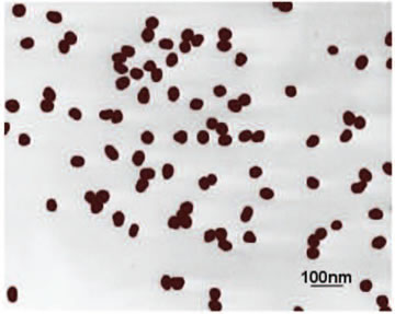
In the development of nanoparticles, there are several conventional particle characterization techniques. Particle imaging techniques using electron microscopy have been widely used to understand the structure and morphology of individual particles in the system (Figure 1). However, several limitations of this technology limit its practical application in routine analysis.
|
Figure 1 From a statistical point of view, in the study of nanoparticle uniformity and aggregation, it is not a good method to rely on electron microscopy for volume-based particle size distribution measurements. |
Electron microscopy is only able to measure a small sample distribution and is based on the number of particle size averages. Colloidal gold products are made up of thousands of particles, so from a statistical point of view, it may not be a good idea to infer the overall uniformity or aggregate concentration of a product based on such data. Even with low amounts of aggregates, clinical outcomes are affected, and this often indicates problems with processing or formulation, so quality control cannot rely solely on electron microscopy. In addition, analysis by electron microscopy is usually time consuming and intense, and is costly and labor intensive. Therefore, a collection technique is needed to supplement the particle size distribution in the entire dispersion based on volume or mass to identify substandard aggregates.
Dynamic Light Scattering (DLS) is a non-invasive technique commonly used to analyze dispersed and colloidal nanoparticles. The DLS technique detects suspended particles with random Brownian motion to obtain fluctuations in the intensity of the scattered light over time. After analyzing these fluctuations in light intensity, the diffusion coefficient can be obtained, and then the particle size is calculated by the Stokes-Einstein equation.
In recent years, some advances in DLS technology have increased the sensitivity and resolution of DLS measurements in the nanoscale range. For example, patented non-invasive backscatter (NIBS) optical instruments are now available for measurement in the dynamic particle size range, with particle diameters ranging from 0.3 nm to 10 microns and solution concentrations ranging from 0.1 ppm to 40% w/v. . Using DLS technology for data collection is fast, does not affect sample recovery, and can play an effective role in both research and quality control.
The following case study explores the advantages of DLS technology for nanoparticle characterization. The experimental data of colloidal gold highlights the differences between the results obtained by DLS technology and electron microscopy, and how to better understand the nanoparticle system.
Case Study: Colloidal Gold Characterization Using DLS Technology
The experiment used a high-level DLS system (Zetasizer Nano S) to measure a colloidal gold sample. All measurements were taken at 25 ° C. The DLS system used a 633 nm laser source and an avalanche photodiode (APD) as the photodetector to detect a scattered light angle of 173 degrees.
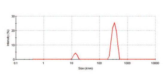
Figure 2 shows the light intensity particle size distribution of a colloidal gold sample. The figure shows the relative percentage of light scattering of particles in different particle size classes. Two different peaks at 13.6 nm and 339 nm, respectively, indicate a bimodal distribution of the particles, indicating the presence of aggregates in the sample.
Â
|
The bimodal intensity particle size distribution of the colloidal gold sample in Figure 2 indicates that there are aggregates in the sample. |
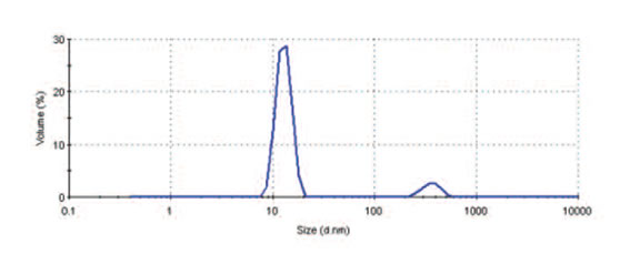
First, from the relative intensity of the peak of the particle size distribution, there appears to be a large number of aggregates in the sample. However, when converting it into a volume-based distribution, as shown in Fig. 2, it is apparent that the concentration of aggregates is actually relatively low. This conversion is performed by intelligent instrument software that uses the Mie scattering theory and the refractive index and absorptivity of the particles. The volumetric particle size distribution shows that in the unit mass, most of the sample particles are small particles of about 13 nm, and the ratio of the original particles to the aggregate is 9:1.
|
Figure 3 shows the volume distribution of colloidal gold samples analyzed by DLS technology. More than 90% of the samples consist of small particles of about 13 nm. |
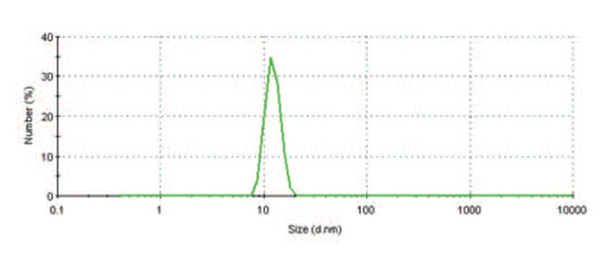
Comparing volume- or light-based particle size measurements with volume-based analytical methods, the value of complementing DLS technology with traditional nanoparticle analysis techniques is found. As shown in Figure 4, the volume-based distribution is converted into a quantity-based distribution. Since the sample contains very few aggregates, the number distribution is a single peak with a peak mean of 12.4 and only the original particles are considered. This result indicates that if the sample is characterized by an electron microscope or the like, most of the particles presented will be small particles, and thus it is difficult to accurately infer the overall concentration of the aggregate.
|
Figure 4 shows the particle size of the number distribution and the unimodal distribution of the original particles. |
The dynamic light scattering resolution is relatively lower than that of electron microscopy. Generally, if the particle diameters differ by a factor of three or more, separate distribution peaks can be obtained by dynamic light scattering techniques. However, through intelligent data interpretation, we can still know the particle size distribution information of quite a lot of samples. For example, a single particle may produce a broad peak when mixed with an aggregate of 2, 3 or 4 particles. Since larger particles scatter most of the light, the aggregate has a greater influence on the peak than the smaller particles. Both the Z-average diameter and the polydispersity index values ​​are sensitive to the presence of aggregates and therefore indicate well the presence of aggregates within the sample. The Z-average diameter is the average of the intensity-weighted hydrodynamic diameters, and the polydispersity index PDI characterizes the particle size distribution width. Both parameters are calculated by the system according to the International Standard for Dynamic Light Scattering ISO22412.
About dynamic light scattering
The development and formulation of nanoparticles requires fast and reliable quality control analysis of the entire sample, while dynamic light scattering provides a solution to meet this need. Through highly statistically significant particle size distribution maps, DLS technology enables users to quickly identify aggregates or substandard particles, which may not be possible if only particle-based particle size measurement techniques are utilized. Today, commercial DLS systems can already use this high-end technology to provide the high sensitivity, high accuracy and high resolution analysis required for nanoparticle uniformity and aggregation research in advanced biomedical applications.
Stephen Ball is the marketing manager for Malvern Instruments' Nanoparticle and Molecular Characterization products. He received a degree in Computer-Aided Chemistry from the University of Surrey, England, where he spent a one-year industrial internship as a research chemist at Dow Chemical in Horgen, Switzerland. Prior to joining Malvern Instruments, he worked as an application chemist at Polymer Laboratories and later as a product manager for light scattering instrumentation at Agilent Technologies.
Digital Imaging Equipment,X-Ray Equipment And Accessories,16 Slice Ct Scan,Ct Scanner
Shanghai Rocatti Biotechnology Co.,Ltd , https://www.ljdmedicals.com


