Operation procedure of enrofloxacin drug residue detector in aquatic products
Enrofloxacin is a chemically synthesized broad-spectrum antibacterial drug and is a representative product of fluoroquinolones. It is widely used in aquaculture and clinical fields.
There are more and more reports on the drug resistance, residual toxicity and adverse reactions of these drugs.
Many countries in the world have developed maximum residue limits for animal foods for this class of drugs.
The detection of enrofloxacin generally uses chromatography as a confirmatory test method; in recent years, immunoassay methods for rapid screening and detection have attracted more and more attention.
This study aimed to establish a one-step ELISA rapid detection method for enrofloxacin residues in aquatic products to further improve the efficiency of rapid screening detection.
A one-step indirect competitive ELISA detection system for enrofloxacin was established by optimizing the traditional two-step indirect competitive ELISA competitive reaction mode, reaction time and washing time. The coupling method was established and optimized by different coupling methods. The method for synthesizing enrofloxacin enzyme-labeled antibody was established. Based on the prepared enzyme-labeled antibody, a one-step direct competition ELISA detection mode was established, and the detection of the spiked aquatic product was verified.
Required equipment and reagents
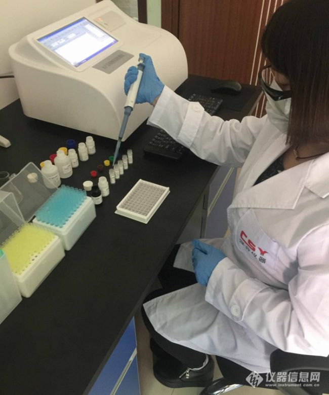
4.1 Instrument: CSY-E96S aquatic product drug residue detector, homogenizer, nitrogen blow dryer, oscillator, centrifuge, graduated pipette, balance (sensitivity 0.01g)
4.2 Micropipette: single channel 20μl-200μl, 100μl-1000μl, multi-channel 300μl
4.3 Reagents: Anhydrous acetonitrile, n-hexane, concentrated HCl
5 sample pretreatment
5.1 Notes before sample processing:
Laboratory equipment must be clean and use disposable tips to avoid contamination and interference with experimental results.
5.2 Dosing:
Solution 1: 0.15 M hydrochloric acid solution 5 ml of concentrated hydrochloric acid was taken and added to deionized water to make up to 400 ml.
Solution 2: Sample extract Amount of 10 ml of 0.15 M hydrochloric acid solution (Liquid 1) was added to 90 ml of anhydrous acetonitrile and mixed well.
Solution 3: complex solution The 5× complex solution was diluted 5 times with deionized water for reconstitution of the sample, and the complex solution was stored at 4 ° C for one month.
5.3 Sample pre-processing steps:
5.3.1 Treatment of animal tissues (chicken, pork, fish, shrimp)
1) Weigh 2.0±0.05g of homogenized tissue sample in a 50ml centrifuge tube;
2) Add 8 ml of sample extract (liquid preparation 2), shake for 5 minutes, and centrifuge at room temperature for 4000 minutes/separate for 10 minutes;
3) Take 2 ml of the clear upper organic phase into a clean and dry 10 ml glass test tube, and blow dry with a nitrogen bath at 50-60 ° C;
4) Add 1 ml of n-hexane, shake for 2 minutes, add 1 ml of the complex solution (dispensing solution 3), shake for 30 seconds, room temperature 4000 rpm / separate the heart for 5 minutes;
5) The upper layer of n-hexane was removed, and 50 μl of the liquid in the lower aqueous phase was taken for analysis.
Sample dilution factor: 2 Detection limit: 0.3ppb
5.3.2 Honey treatment method
1) Take 1.0±0.05g honey into a 50ml polystyrene centrifuge tube, add 6ml sample extract (solution 2), shake for 5 minutes, and make it fully dissolved;
2) Add 3 ml of the complex solution (dispensing solution 3), add 11 ml of dichloromethane, shake for 5 minutes, room temperature 4000 rpm, and centrifuge for 5 minutes;
3) Aspirate the upper layer, remove 8 ml of the lower organic phase into a desiccator, and blow dry with a nitrogen bath at 50-60 ° C;
4) Dissolve the dried residue with 1 ml of reconstituted working solution, add 1 ml of n-hexane and mix for 30 seconds, room temperature 3000 rpm or more, and centrifuge for 5 minutes;
5) Remove the upper layer and remove 50 μl of the liquid for analysis.
Sample dilution factor: 2 Detection limit: 0.4ppb
5.3.3 Milk treatment method:
1) Mix 25 μl of the sample solution with 475 μl of the complex solution (dispensing solution 3), shake for 1 minute, and dissolve it sufficiently;
2) Take 50 μl of liquid for analysis.
Sample dilution factor: 20 detection limit: 3ppb
5.3.4 Milk powder treatment method:
1) Weigh 0.5 ± 0.02 g of homogenate into a 10 ml polystyrene centrifuge tube, add 5 ml of deionized water, shake to make it fully dissolved;
2) 100 μl of the sample solution was mixed with 400 μl of the complex solution (dispensing solution 3), and shaken for 1 minute;
3) Take 50 μl of liquid for analysis.
Sample dilution factor: 50 detection limit: 6ppb
5.3.5 Egg processing method:
1) Weigh 1.0 ± 0.02 g of homogenate into a 10 ml polystyrene centrifuge tube, add 5 ml of deionized water, shake to make it fully soluble;
2) 100 μl of the sample solution was mixed with 400 μl of the complex solution (dispensing solution 3), and shaken for 1 minute;
3) Take 50 μl of liquid for analysis.
Sample dilution factor: 30 detection limit: 3ppb
6 Enzyme-linked immunoassay procedure Take the required reagents from the 4 ° C refrigerated environment and equilibrate at room temperature for more than 30 min. When the washing liquid is refrigerated, there may be crystallization to return to room temperature to fully dissolve. Each liquid reagent must be shaken before use. uniform. Remove the required number of microplates and frames, place the unused microplates in a ziplock bag and store at 2-8 °C.
Before the start of the experiment, the 20X concentrated washing solution was diluted with 20 times of deionized water into a working washing solution.
6.1 No.: The micropores corresponding to the sample and the standard are numbered sequentially. Each sample is parallel to the standard 2 holes, and the position of the standard hole and the sample hole is recorded.
6.2 Addition reaction: Add standard or sample 50μl/well to each microwell, then add 50μl/well of enzyme label, then add 50μl/well of antibody working solution, seal the plate with cover membrane, and gently shake 5 Mix in seconds and react at 25 ° C for 45 minutes.
6.3 Washing: Carefully uncover the cover film, dry the liquid in the well, and wash it thoroughly with the working washing liquid 250μl/well 5 times, each time interval 30 seconds, pat dry with absorbent paper (bubbles that have not been removed after pat drying) A clean gun puncture).
6.4 Color development: Add 50 μl of substrate solution A to each well, add 50 μl of substrate solution B, mix gently by shaking for 5 seconds, and develop color for 15 minutes at 25 ° C in the dark.
6.5 End: Add 50 μl of stop solution to each well, mix gently by shaking, and stop the reaction.
6.6 Measurement of absorbance: The absorbance value of each well was measured at 450 nm using a microplate reader (double wavelength 450/630 nm is recommended). The assay should be completed within 10 minutes of terminating the reaction.
7 Results analysis
7.1 Calculation of Percent Absorbance The percent absorbance of the standard or sample is equal to the average of the absorbance values ​​of the standard or sample (double well) divided by the absorbance of the first standard (0 ppb), multiplied by 100%, that is, the absorbance value (%) = A × 100%
A0
A—the average absorbance value of the standard solution or sample solution
Average absorbance value of A0—0ppb standard solution
7.2 Drawing and calculation of the standard curve The percent absorbance of the standard solution is plotted on the ordinate, and the logarithm of the corresponding standard solution concentration (ppb) is plotted on the abscissa. The semi-logarithmic plot of the standard solution is plotted. Substituting the percent absorbance of the sample into the standard curve, reading the concentration corresponding to the sample from the standard curve, and multiplying the corresponding dilution factor is the actual concentration of the analyte in the sample.
If the kit professional analysis software is used for calculation, it is more convenient for accurate and rapid analysis of a large number of samples. (Welcome to call)
8 Precautions
8.1 Room temperature below 25 ° C or reagents and samples not returned to room temperature (25 ° C) will result in low OD values ​​for all standards.
8.2 If the plate hole is dry during the washing process, the standard curve will not be linear and the repeatability is not good. Therefore, the next step should be taken immediately after the plate is patted dry.
8.3 The mixing should be uniform, the washing should be thorough, and the reproducibility in the ELISA analysis depends largely on the consistency of the washing.
8.4 Seal the microplate with a cover film during all incubations to avoid light exposure.
8.5 Do not use kits that have expired. Do not exchange reagents from different batches.
8.6 If any color of the coloring solution indicates deterioration, it should be discarded. A 0 standard absorbance value of less than 0.5 units (A450nm < 0.5) indicates that the reagent may deteriorate.
8.7 The reaction stop solution is corrosive and avoids contact with the skin.
Http://
NATURAL is one of the most professional Animal Feed Ingredients manufacturers and suppliers.
Animal feed ingredients main function including:Improve animal immunity, Feed additives, The efficacy of aquaculture, Promote animal growth
Main product: Astragalus polysaccharides, Curcumin, Lentinan, Yucca, Rhubarb extract, Chlorella tablets, Dihydromyricetin and 5-Aminolevulinic acid hydrochloride
Welcome to contact us.
Animal Feed ingredients, Animal Feed Raw Ingredients, Natural Field Animal Feed ingredients,Factory feed raw material
Xi'an Natural Field Bio-Technique Co., Ltd. , https://www.natural-field.com